Iridology International

Home / Programs

Registered Iridologist Diploma (R.Irid.)

A complete, graded introduction to the iridological sciences. It runs from established principles to current theory and research, and draws on Jensen, Vida-Deck, Günther, Roberts, Bourdiol, Angerer, Velchover and Jausas.

  • 5 modulesof online lessons
  • 20+ hoursof study material
  • 75% to passeach module exam
  • 12 monthsto complete

About the program

The R.Irid. program is Iridology International's core diploma. You study online, in order, at your own pace, and a graded exam closes each module. Along the way you learn to examine an iris with a magnifier, a camera, a digital iriscope and software.

What sets it apart is breadth. Most English-language courses teach one American chart. This one sets Jensen's work beside the European and Russian research that followed it, so you understand where the charts agree, where they differ, and why.

What you'll learn

  • Iris reading technique with a magnifier, camera, digital iriscope and software
  • How American (Jensen) iridology compares with current European and Asian research
  • How structural, reflex and pigment signs point to where a process is located
  • Reading the collarette (autonomic nerve wreath) for inherent weakness
  • Changes in pupil colour, size, shape, position, equality and light reaction, and their significance
  • What the conjunctival vessels, lens and cornea add to an iris assessment
  • Hereditary iris colour, toxic and drug-related dyschromia, and pigment spots
  • The constitutions and their sub-types
  • Completing a full iris analysis from start to finish
  • The central hypotheses of iridology, in historical and current context

Syllabus

1

Module 1: Introduction to Iridology

Where iridology came from, how it is studied, and the vocabulary used for the rest of the program.

  1. Introduction to IridologyThe roots of iridology, from findings in Tutankhamun's tomb to current technology and recent clinical studies.
  2. Clinical Study ApproachesEmpirical investigations, the central hypotheses of iridology, and the statistical concepts needed to read a study critically.
  3. Iridological Concepts and ScreeningsThe classes of structural change seen in the iris and how they are screened.
  4. TerminologyThe program glossary: the terms used in every lesson that follows.
  5. Ancient Symbols of the EyeAn optional lesson on the eye in ancient history and symbolism.
2

Module 2: Structural, Reflex and Pigment Signs of the Eye

The core of the program. How structural, reflex and pigment signs indicate where a process is located and, as far as iridology allows, its mechanism, stage, character and severity.

  1. Anatomy of the EyeAnatomical fundamentals, histology and anomalies of the human eye.
  2. LacunaeHow lacunae are assessed by size, depth, shape, colour, location and the state of their edges, and how their relationship to other organs and to the autonomic nerve wreath shapes an assessment.
  3. Topolabile and Topostabile SignsSigns whose meaning depends on their location in the iris, and signs whose meaning does not, and why the difference matters.
  4. Reflex SignsVascular signs in the iris. Radial vascular signs are phenotypic, and are read as irritation or congestion in the associated organs.
  5. Crypts and DefectsSmall, dark, often black lacunae. When very small and dark they are called defect signs: a loss of integrity of the iris stroma that is genetically determined.
  6. Iris PigmentationTelling the inherited base colour, governed by regulator genes, apart from foreign pigment of pathophysiological origin.
  7. TransversalsAmong the most important focal signs in the iris. How true transversals are read as a tendency toward inflammation, injury, irritation, pain and tissue degeneration.
  8. Relief of the Iris SurfaceThe iris surface is never flat: it is a landscape of elevations and depressions. Reading the relief gives an impression of the body's reserve and self-protective capacity.
  9. The CollaretteThe autonomic nerve wreath, one of the most important landmarks an iridologist reads, and its role as a marker for the gastrointestinal tract and the autonomic nervous system.
  10. Toxic and Dystrophic Changes of the IrisToxic radii, white radii, slagging, the dystrophic rim, the lymphatic rosary and the senile arc, and their proposed link to the five eliminative systems: lungs, bowel, kidneys, skin and lymph.
  11. Heterochromia, Dyschromia and PathochromiaComplete (true and false) and partial (central and sectoral) heterochromia, and how central heterochromia shows as a colour difference between the pupillary and ciliary zones.
  12. Adaptive Rings and ArcsOphthalmology treats contraction rings as simple folds from the eye's neuromotor apparatus. Iridologists consider other factors too, including the person's emotional state.
  13. Axis SignsGroups of lacunae, crypts, radial furrows, pigment and collarette signs that line up in specific locations, and what they add to an assessment.
  14. Conjunctival IndicatorsThe bulbar conjunctiva does not repeat the iris examination: it adds new information. Why and how to include it.

3

Module 3: Constitutional Classification

The three main constitutions (lymphatic, haematogenic and biliary or mixed) with their sub-types and syndromes, so common variations in physiology and predisposition can be recognised.

  1. The Lymphatic ConstitutionThe lymphatic constitution and its sub-types.
  2. The Biliary or Mixed ConstitutionThe classic biliary constitution and its sub-types.
  3. The Haematogenic ConstitutionThe haematogenic constitution and its sub-types.
  4. Pathological Constitutions and PredispositionsConstitutional patterns associated with specific predispositions.
  5. Constitutional SyndromesHow constitutional signs combine into recognisable syndromes.
  6. The Three Pre-Cancerous Constitutions According to BroyJosef Broy's classification and how it is used in constitutional research.
  7. Constitutional Modification: a Homeopathic GuideA reference to the homeopathic elements used in constitutional modification research.
4

Module 4: The Pupils

Pupil tone reflects the central and autonomic nervous systems. Changes in pupil colour, size, shape, position, equality and light reaction all carry clinical significance.

  1. The PupilsWhy pupil reflexes matter so much in neurological assessment, and what to look for.
  2. Pupil DeformationsTelling a deformation caused by local disease of the iris apart from one that develops reflexively from disease of the internal organs or central nervous system.
  3. Pupil DecentrationNormally the pupil sits centrally or slightly inward and downward. How shifts in position reflect the balance between the sympathetic and parasympathetic nervous systems.
  4. The Pupillary MarginThe pigment fringe at the inner edge of the iris. How to distinguish the six main types of pupillary margin sign, and why four of them are linked to specific conditions.
5

Module 5: Iridodiagnosis: Examination Procedures

Putting it into practice: examination technique, equipment, iridology charts, and assessing an iris with the included software.

  1. Examination ProceduresAn assessment is built on visual examination of the iris and its signs. This lesson covers the procedures needed to do that accurately.
  2. Iridology ChartsWhy there are so many iris charts, and how chart development has progressed from Jensen to the European and Russian schools.
  3. Installing the Iridology Analysis ToolkitInstalling the included software and importing your first eye images.
  4. Assessing an Iris with the ToolkitA walk through each section of a software assessment.
  5. Reading the Assessment ResultsThe terminology and results the software produces, and how to interpret them.

Exams and diploma

Each module has a practice exam and a graded exam, both taken online. You need 75% or more to pass. If you score lower, you retake the exam with a different set of questions drawn from the question bank. The program ends with a 50-question final exam.

Every exam is reviewed by Iridology International, and your results are emailed to you. When you have passed every module and the final, you receive the Registered Iridologist (R.Irid.) diploma. Students with consistently high marks graduate with honours. You can request an academic transcript at any time.

What's included

  • The online program at students.iridologyinternational.com, with all lessons and study materials
  • Iridology Analysis Toolkit, an unlimited license with lifetime updates ($449 if bought alone)
  • Iridology Research, lifetime membership ($495 if bought alone)
  • All module exams, the final exam and your diploma
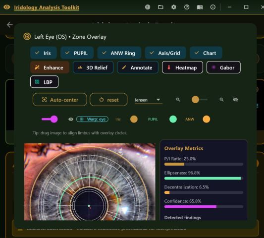
Toolkit zone overlay on a left iris, with toggles for pupil, collarette ring, chart, enhancement, 3D relief and heatmap, and overlay metrics.
The Toolkit's zone overlay. Module 5 teaches you to install it and read its results.

An iriscope isn't included and isn't required: a 4× magnifying glass is enough for the course. For iris photography, the Toolkit is also sold with a Dino-Lite iriscope.

Requirements

Applicants need a working knowledge of medical terminology, human anatomy and physiology. If you don't have that background yet, take an introductory course in those subjects before you start, or alongside Module 1.

You'll need a computer or tablet with internet access. The Toolkit runs on Windows 10 or later, Android, iOS and macOS.

Tuition

Please note. Iridology is a complementary assessment method, not a substitute for medical diagnosis. The R.Irid. is a professional credential from Iridology International, not a license to practise medicine. No iridology school currently holds recognised accreditation.